| |
 |
| |
| |
| |
| 194 SLICE CARDIAC CT |
| |
| |
|
|
| |
iScan Imaging Centre is equipped with Siemens SOMATOM Sensation CT Scanner,
which is an excellent time tested and tried top-of-the line ultrafast 194 slice CT scanner. It is an extremely fast multidetector
scanner with tube rotation of 330 ms and high temporal resolution of 165 ms, optimum for CT Coronary Angiography.
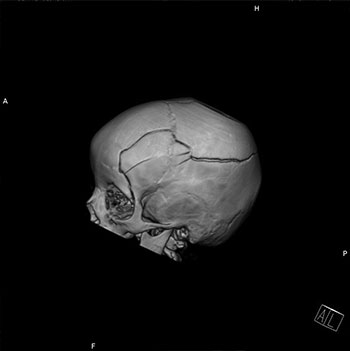
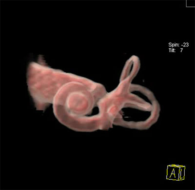
It has high-resolution image capability, with the ability to create brilliant 2D and 3D images.
CT means 'Computed Tomography'. CT Scanners have revolutionary evolved from the conventional slice-to-slice scan to spiral/ helical scan and then to multidetector/
multislice scan,which are capable of acquiring multiple sections per second significantly reducing scanning time and improving patient compliance.
They use X-ray tube and multiple rows of detectors which rotate around the patient providing diagnostic images through body volume, with isotropic
multiplanar imaging. The scanner gives high-resolution images of any part of the body, but is especially excellent for cardiovascular, lung and abdominal imaging.
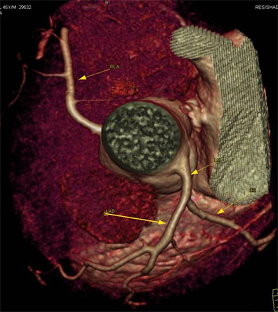
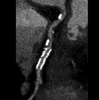
Somatom Sensation is excellent for cardiac imaging, where "frozen" images of the heart can be obtained with optimal evaluation of the coronary arteries
and cardiac function within 10 sec. This may even obviate or delay the need for routine angiography.
CT Scan is by far the best method of detailed visualization of the lungs, especially for characterization of interstitial and diffuse lung diseases.
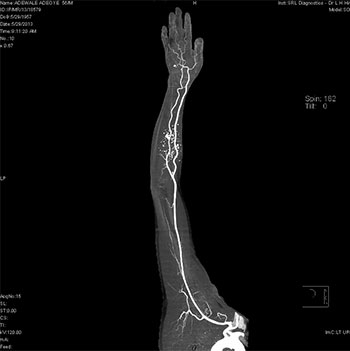
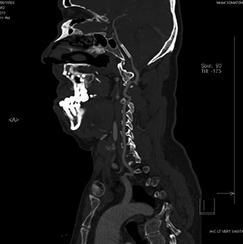
It is also the gold standard for the detection of renal stones. Multislice CT Angiography has become a gold-standard and replaced diagnostic
DSA in many conditions and can be combined with sectional imaging. It provides extremely fast and high-resolution images of the abdomen,
including bowel abnormalities and can be performed even in uncooperative patients, which is difficult on MRI. CT scan is safe , excellent
and fast method of diagnosing and staging cancers,and combined whole body PET-CT is gold standard in oncology practice. Procedures
like CT-guided biopsies, aspirations procedures and radiofrequency ablations can also be safely performed with good patient compliance.
CT can be used in patients who cannot undergo MRI e.g. patients with pacemakers, cochlear implants and aneurysm clips. It can also be used in the presence of
an implant, where MRI can leave significantly degrading artefacts, precluding proper visualization of the part.
Radiation hazards warrant judicious use of CT in pediatric and pregnant patients, but can be minimized with patient and body-specific protocols and dose reductions.
Non-ionic contrast used in CT can have mild and rare anaphylactic reactions due to iodine content and should be judiciously used in patients with impaired renal function. |
|
| |
|
|
|
|
|
|