|
iScan Imaging Centre is equipped with Siemens MAGNETOM Spectra 3 Tesla MRI, which is the first of its kind in Mumbai. It is the first private diagnostic centre in South Mumbai equipped with a 3 Tesla Magnet. It provides unprecedented access to a new level of diagnostics with excellent resolution system, unchallanged image quality and all the advanced applications in MRI.

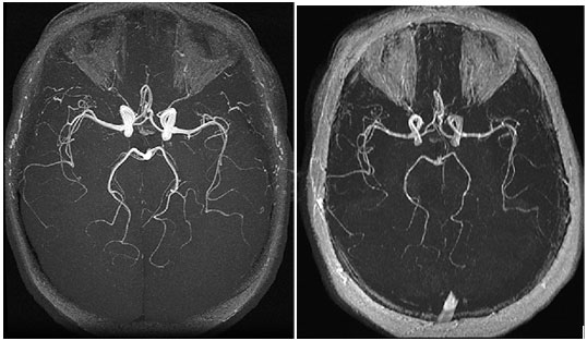
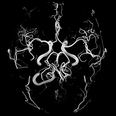
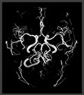
Normal arterial circulation: 3D TOF study at 3.0 T (a) and 1.5 T (b). Vascular conspicuity and background are greater in a than in b , resulting in greater vessel-tissue contrast
MRI means 'Magnetic Resonance Imaging'. It's a diagnostic imaging technique using a strong magnetic field to produce high-quality pictures of soft tissue inside your body, for example nerves, muscles and blood vessels. It does not involve radiation and nothing has to be inserted into your body, in short – MRI is a noninvasive diagnostic tool that allows your physician to see pictures of the inside of your body without surgery and without any harmful effects on the body.
MRI scan is able to provide clear pictures of parts of the body that are surrounded by bone tissue, so the technique is useful when examining the brain and spinal cord. MRI scan gives very detailed pictures when it comes to finding tumours (benign or malignant abnormal growths) in the brain. If a tumour is present the scan can also be used to find out if it has spread into nearby brain tissue. The technique also allows us to focus on other details in the brain. For example, it makes it possible to see the strands of abnormal tissue that occur if someone has multiple sclerosis. It is possible to see a change occurring when there is bleeding in the brain, or find out if the brain tissue has suffered lack of oxygen after a stroke.The MRI scan is also able to show both the heart and the large blood vessels in the surrounding tissue. This makes it possible to detect heart defects that have been building up since birth, as well as changes in the thickness of the muscles around the heart following a heart attack. The method can also be used to examine the joints, spine and sometimes the soft parts of your body such as the liver, kidneys and spleen. Also with advanced applications, the metabolic and perfusion data can be obtained, which will further help in disease characterization.
MRI is different from CT Scan, which is also a widely used diagnostic test. There is no ionizing radiation (X-rays) involved in producing an MRI scan. MRI scans are generally more detailed, too. The difference between normal and abnormal tissue is often clearer on the MRI scan than on the CT scan.
With nearly two times more signal than 1.5T MR, 3Tesla MRI provide significant improvement in temporal and spatial resolution or to apply imaging techniques that are not otherwise clinically feasible, due to long scanning times. Routine MR Brain can be performed in a patient from intensive care or a child in upto 7 minutes.
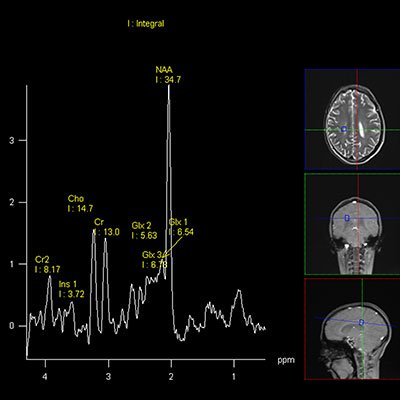
MRI on 3Tesla is twice as fast as 1.5T with the same SNR or provides twice as much resolution, or a combination of the two. It results in a definative improved diagnostic sensitivity and specificity. The finer spatial resolution of 3T helps conduct high-resolution inflow MR angiography, bringing it close to the resolution of DSA and enhance the sensitivity of diffusion studies. MR Spectroscopy on 3T has more signal and better defined metabolic peaks, making it and DTI to be performed in routine imaging time.
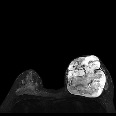
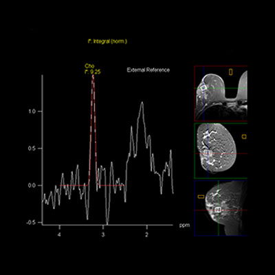
Speed at 3T is particularly helpful in performing Cardiac MR and the most promising application is in myocardial perfusion.MR at 3T seems to be ideally suited to Breast MR because it has enough signal to use a higher imaging matrix and obtain contrast from lesions before normal breast tissue enhances in the early postcontrast phase. In breast imaging, extremely high spatial resolution is needed to depict the small morphological details of cancers. Arterial spin labeling (ASL) to measure blood flow in the brain or heart on 3T MRI, can be performed in only about four minutes due to the speed and outstanding resolution.
In Body imaging, 3T MR helps in detection of small and ultrasmall lesions of the liver due to improved spatial resolution which aids in planning radio-frequency ablation and stereotactic radiosurgery. The combined high spatial resolution DCE &T2W MRI at 3T provides improved assessment and high staging accuracy of prostate cancer. The 3T resolution & contrast is far superior in evaluation of uterine & cervical pathologies than at 1.5T.
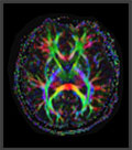
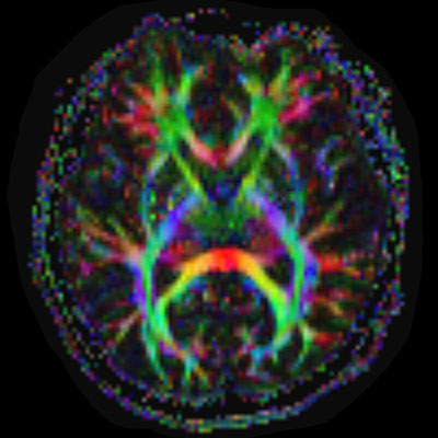
3T - Head EPI DTI FA map colored
|
|
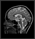
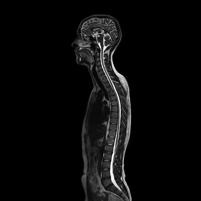
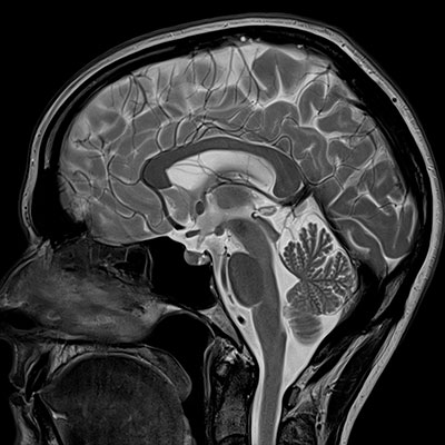
01j_C05_Head_T2_TSE_sag_512
|
|
3T_Fusion_MapIt_sag
|
|
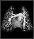
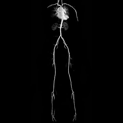
3T Angio
|
|
|
|
|
|
|
|
3T - Head EPI DTI FA map colored
|
|
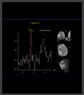
3T Breast Spectro
|
|
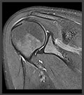
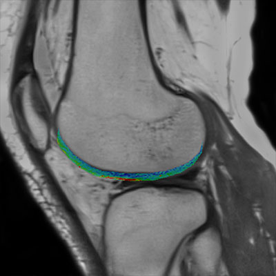
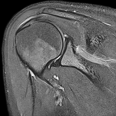
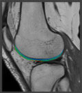
3T Shoulder
|
|
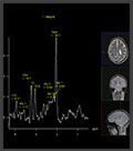
3T_Spectro_Spectrum
|
|
In Orthopedic imaging, 3T MR provides double the inplane resolution and upto 4 times the speed on multiple-average scans. Along with dedicated Ipat-compatible extremity, optimised fat saturation can be obtained with extremely high resolution, improving patient comfort and throughput. Improved resolution aids better visualization of the articular cartilage of the hip/ acetabulum, talar dome and the wrist and small structures like labrum, TFCC, nerves and tiny ligaments. Cartilage MapIT allows the early detection of osteoarthritis and cartilage transplant evaluation.
The Siemens Spectra is a true 24 RFchannel system for upto 120 coil elements with ultra light-weight high density coil array covering from head-to-toe. It is provided with patient-table-integrated 24-element Spine coil, 18-channel knee coil and 16-channel Head/ Shoulder/ Foot coils with capability of coil combinations. It has a bore of 60 cm with the shortest system length of only 173 cm for less patient claustrophobia. It has Tim 4G and Tim TX TrueForm with automated Dot Engine for enhanced patient comfort. |
|